Welcome to The Jabbarzadeh Group.

301 Main St
Columbia, SC 29201

803-777-3297
ehsan@sc.edu

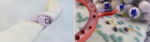
Cell morphodynamics and interactions with microenvironment

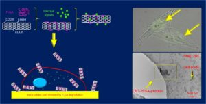
Human pluripotent stem cells (hPSCs) offer a promising tool for regenerative therapies because of their unique capability to self-renew, proliferate nearly indefinitely, and rise to various cell types. In the regeneration process, the temporary extracellular matrix (ECM) provides multiple signals to hPSCs and guides the process of new matrix formation. Fundamental questions on how hPSCs sense and respond to the cues from surrounding environment await further exploration. A main hurdle facing the studies of hPSC-ECM interactions lies in limitation of hPSCs culture techniques which use clumps of cells from dissected colonies. In these culture methods, hPSCs unavoidably pass through the stage of spontaneous differentiation to all three embryonic germ layers before the generation of the desired tissue-specific cell type. The goal of this study is to generate and characterize a monolayer single-cell culture model that allows for the investigation of biological phenomena associated with hPSC lineage specification. Specifically, we seek to (1) leverage micropatterning approaches to control cell shape and orientation, and activation of polarity proteins in hPSCs at a single cell level, and (2) quantitatively map the contribution of cell shape to germ layer specification of hPSCs.


Picture1pic1

Representative Publications:

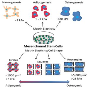
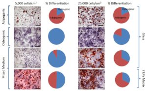
G. Harris, M. E. Piroli, and E. Jabbarzadeh, “Deconstructing the effects of matrix elasticity and geometry in mesenchymal stem cell lineage commitment,” Advanced Functional Materials 14: 2396-2403 (2014). Download PDF

G. Harris, T. Shazly, and E. Jabbarzadeh, “Deciphering the combinatorial roles of geometric, mechanical, and adhesion cues in regulation of cell spreading,” PLoS One 8(11) e81113 (2013). Download PDF


Vascularization of biomaterials for bone repair

Vascularization of biomaterials is a major challenge in tissue engineering and thus, our success in creating off-the-shelf products has been limited mostly to thin avascular tissues such as skin and cartilage. The primary function of vasculature is to facilitate the transport of nutrient and oxygen to the cells invading from the host tissue and cells transplanted in the scaffolds. Insufficient neovascularization can result in the formation of fibrous capsule surrounding the implants and prevention of biochemical transport into their interior. Our lab is interested in developing new strategies to promote vascular infiltration in porous scaffolds. We set out to harness the wound healing potential of macrophages and human mesenchymal stem cells (hMSCs) to enhance directed angiogenesis and bone formation in three dimensional (3D) PLGA sintered microsphere scaffolds for osseous repair.Towards this goal, we will identify the nexus of antiinflammatory natural compounds and oxygen signaling that promote wound-healing macrophage phenotype, and enhance osteogenic and angiogenic potential of hMSCs. Our effort is motivated by the potential of (1) natural antioxidants to modulate inflammation in a number of diseases including cancer and cardiovascular disorders, and (2) ischemic pre-conditioning of stem cells to drive physiological release of angiogenic factors. The figure below summarizes the vision underlying our work.

research

Representative Publications:

K. E. Rutledge, Q. Cheng and E. Jabbarzadeh, “Modulation of inflammatory response and induction of bone formation based on combinatorial effects of resveratrol,” Journal of Nanomedicine and Nanotechnology, 7(1):1-10 (2016). Download PDF

M. L. Skiles, B. Hanna, L. Rucker, A. Tripton, A. Brougham-Cook, E. Jabbarzadeh, and J. O. Blanchette*, “ASC Spheroid Geometry and Culture Oxygenation Differentially Impact Induction of Preangiogenic Behaviors in Endothelial Cells,” Cell Transplantation 24(11): 2323-35 (2015). Download PDF

E. Jabbarzadeh, T. Starnes, Y. M. Khan, T. Jiang, A. J. Wirtel, M. Deng, Q. Lv, L. S. Nair, S. B. Doty and C. T. Laurencin, “Induction of angiogenesis in tissue engineered scaffolds designed for bone repair: A combined gene therapy-cell transplantation approach,” Proceedings of the National Academy of Sciences 105:11099-11104 (2008). Download PDF

Carbon nanotube based gene delivery

Viral vectors are efficient in both delivery and transduction of cells; however, there are concerns that the viruses are sourced from lethal diseases such as human immunodeficiency virus (HIV) and human T-cell lymphotropic virus (HTLV). A safe, inexpensive, and effective vector has yet to be fabricated for use in gene therapy. Carbon nanotubes have received an overwhelming amount of support and enthusiasm in biomedical research for their ability to shuttle biological molecules, ranging from small drug molecules to biomacromolecules such as proteins, DNA and RNA into different types of cells via endocytosis. Nevertheless, health concerns and intrinsic toxicity issues have hampered the practical application of using inorganic carbon nanotubes in gene therapy. Our lab is interested in design of functionalization approaches to reduce the toxicity while maximizing the ability to target cells and release multiple factors. The figure below demonstrates our strategy to engineer poly(lactide acid-co-glycolide acid) wrapped carbon nanotubes used to deliver pro-apoptotic molecules with a controlled release profile.

picture1
Representative Publication:
  • Q. Cheng, G. Harris, and E. Jabbarzadeh, “PLGA-Carbon Nanotube Conjugates for Intercellular Delivery of Caspase-3 into Osteosarcoma Cells,” PLoS One 8(12) e81947 (2013). Download PDF

Have questions for our research team, or wish to get in touch?

Contact Us